

Magnetoencephalography Center
Through the generous philanthropic gift of Jack H. Miller, we are proud to provide magnetoencephalography (MEG) brain mapping for pediatric and adult epilepsy and brain surgery patients.
MEG, is one of the world’s most advanced, non-invasive brain mapping technologies available used to precisely locate sources of epilepsy and greatly improve the quality and effectiveness of pre-surgical evaluations. The MEG also localizes special parts of the brain that a surgeon would like to avoid, such as areas that control movement, vision and language.
A premier center of excellence
We are a premier center of excellence and innovation with the only MEG available in West Michigan. The MEG is located at Helen DeVos Children’s Hospital, the only children’s hospital in Michigan offering this technology.
Paul Ferrari, PhD, MEG technical director, discusses how magnetoencephalography works to create a map of the brain.
Your journey with MEG

Getting started
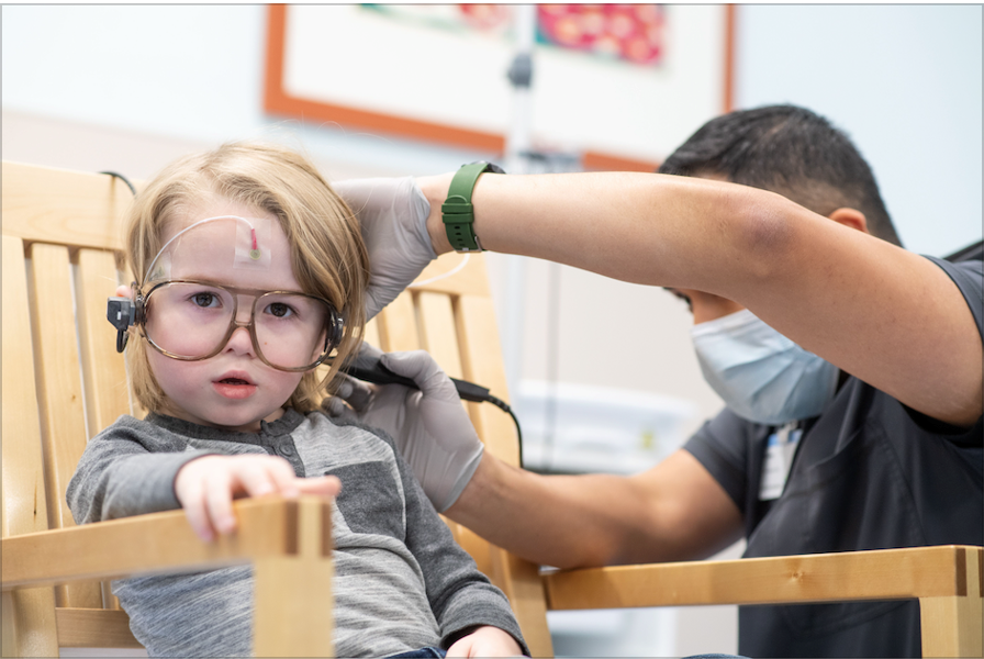
First, the MEG tech will attach a combination of 5 head positioning coils and 22 EEG electrodes to the patient’s head. It may feel sticky, but they are not painful. A member of our child life team can be available to help comfort and distract your child during this process.
Creating coordinates
Next, the MEG tech digitizes the placement of each coil and electrode. The special glasses have a sensor that communicates with a stylus to give 3D coordinates for every coil and electrode. This creates a very precise model of the patient’s head.
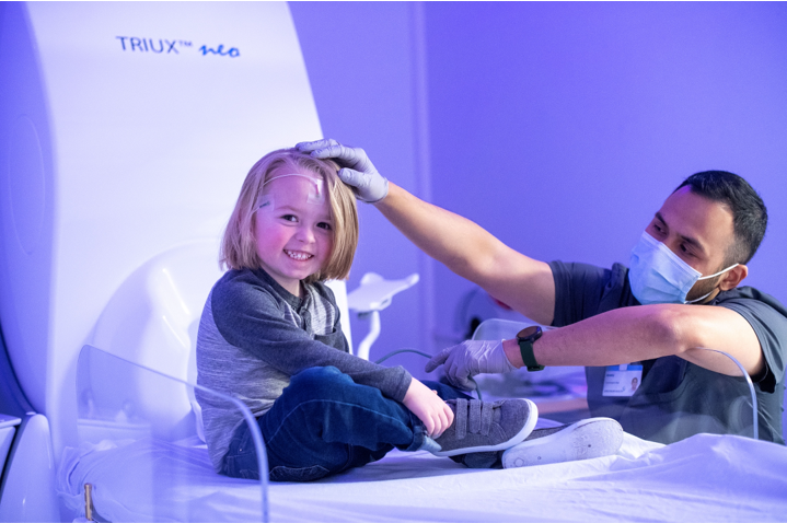
The MEG exam
Then, the patient will go into the MEG room for the actual exam. Many tests will be completed while the patient is awake, including listening for different sounds, feeling different touches or seeing pictures. Most patients will be asked to sleep for part of the exam. It is very important for the patient to keep their head very still throughout the exam.
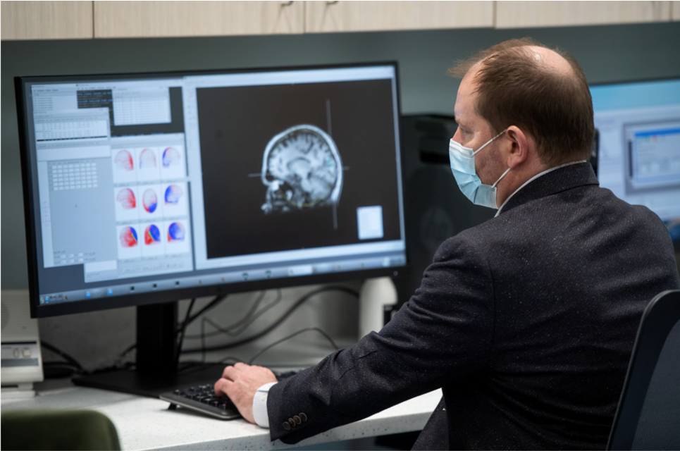
Complete brain map
When the exam is complete, your doctors and surgeons will have a complete map of the brain to prepare for surgery or create a treatment plan.
Advancing neuroscience research
In addition to clinical care, the MEG is one of the most advanced tools for neuroscience research. Our team of neuroscience experts is leading cutting-edge applications for pre-surgical brain mapping and discovering new insights into brain disorders. These areas include epilepsy, neurodevelopmental disorders, stroke and traumatic brain Injury, neuromotor rehabilitation, autism, and also early dedication of Alzheimer’s disease and dementia.
Made possible through generosity
The Magnetoencephalography Center was made possible by Jack Miller. Jack, who served as CEO and president of Howard Miller Company for 48 years, has been a leader in business and philanthropy, supporting a broad scope of initiatives that make West Michigan a better place to live and work. In making this gift, Jack said, “Kindness, compassion, and support for the entire family are vital to the healing process, and Spectrum Health and Helen DeVos Children’s Hospital are filled with hope and healing. I am grateful to be part of a community that is supportive of the health and well-being of future generations and proud to partner with Spectrum Health and Helen DeVos Children’s Hospital in reimagining neuroscience care with the latest in advanced medical technology.
Meet our team


Technical Director
Paul Ferrari, PhD
MEG Technician
Jeremy Gurumendi, R. EEG T
Frequently asked questions

Magnetoencephalography, or MEG, measures the magnetic fields produced by electrical currents occurring naturally in the brain. The measurements create a comprehensive “map” of the brain that pinpoints sources of seizures as well as other essential brain regions and is instrumental in determining the most effective treatment and surgical options.
Benefits:
- Non-invasive. No injections, dyes, radiation or magnetic fields means that studies can be repeated, if needed, without harm or risk.
- Localizes activity in the brain, down to the millimeter.
- Used alongside other diagnostic tools and tests, such as MRI and EEG, to accurately plan for precise surgical procedures.
- For epilepsy, brain tumors and other neurologic conditions.
- Ask your provider about a MEG exam referral.
- After your referral is received by our team, the MEG coordinator will call you for an initial screening and to schedule an appointment. You will also be contacted prior to the exam to make sure preparations are all set. If you or your child will be having sedation for the exam, the sedation team will also contact you.
- If you have any questions about your MEG referral, please call us at 616.486.9995.
- The MEG is totally quiet and does not generate any radiation or magnetic fields. You or your child will lie very still on a bed and wear a special sensing helmet. Your care team will always be able to communicate with and see you.
- Most patients will be asked to sleep for part of the exam. Many tests will be completed while the patient is awake, including listening for different sounds, feeling different touches or seeing pictures. Other tests may involve moving parts of the body, making decisions or answering questions.
Before the exam:
- For epilepsy patients, please limit your sleep to 4 hours or less the night before the exam. We recommend sleeping midnight to 4 am.
- Children less than 5 years of age will be given different instructions.
- Please avoid food or drink with high sugar or caffeine content the evening before or the day of the exam.
Day of the exam:
- Please take medications as prescribed
- Do not wear hair products or any form of cosmetics/make-up
- Please remove any metallic nail polish and all removable piercings
- Epilepsy patients please bring rescue medications
- Check in at your designated location
- You or your child will be asked to remove all metal objects from your body and wear hospital scrubs. There is a secure locker for your clothes and valuables, if needed.
- Epilepsy patients will have an EEG prepared and recorded along with the MEG exam.
Pediatric patients:
- Go to the Helen DeVos Children’s Hospital lobby on the first floor, get your patient badge and take elevators to Level C for check in. The MEG technician will come to get you at your check in area and bring you and your child to the MEG suite on Level-D.
Adult patients:
- Go to the Butterworth Hospital main lobby, get your patient badge, then take the F elevator to 2W to check in. The MEG technician will come to get you at your check in area and bring you to the MEG suite on Level-D.
Depending on how many tests your doctor orders, and if you are having sedation, the exam can take anywhere from two to four hours, including preparations.
Parents and guardians will be allowed to go in the MEG room with your child to help prepare and settle them before the exam, and then will be escorted to the waiting room for the duration of the exam. In some cases, a parent or guardian may need to stay in the MEG suite.
- When the exam is finished, the MEG technician will remove EEG electrodes if they were used. You or your child will change back into his/her clothes, and you will be escorted back to the check-in area.
- The MEG results will be reported directly to your referring physician, usually within two weeks.
Provider Referrals
Schedule a Consultation
Our specialists are ready to serve your child and your family.